|
In accordance with the Inter-granular Pressure Solution (IPS) concept, every grain to grain contact subjected to large enough compressive stress is a potential micro pressure solution location. Another way of putting it: Grain boundaries in rocks under compression act similar to the role of of the so-called 'Griffith Flaws' leading to the initiation and propagation of joints. Here, IPSs along adjacent grains link or coalesce to form a through-going pressure solution seam. Thus what appears to be a single macroscopic pressure solution structure in rock has many intertwined IPSs as shown in Figure 1 and Figure 2. It is therefore an innate property of the pressure solution seams identifiable by naked eyes to have a zonal nature beyond the initial IPS between favorably oriented point contacts of the adjacent grains.
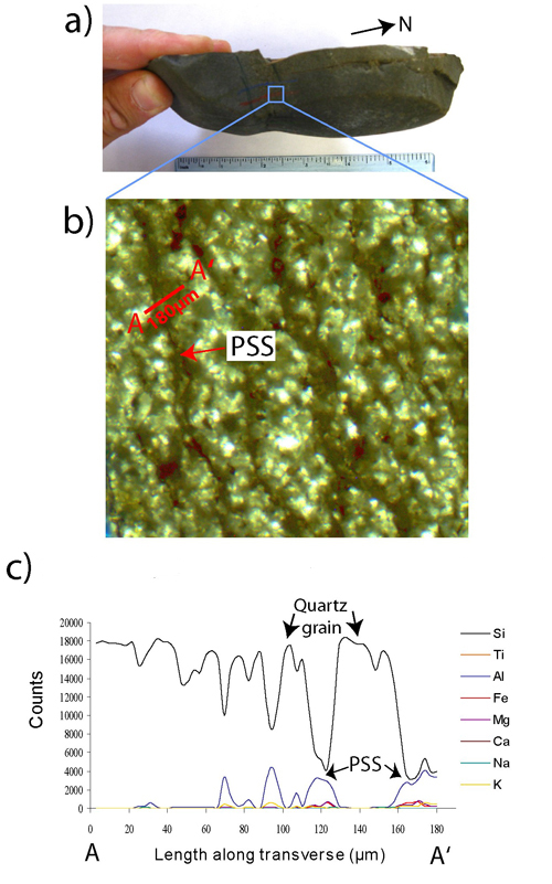
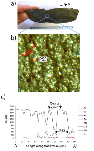 | | Figure 1. Bed-perpendicular pressure solution seams in the Ross Sandstone exposed at Loop Head Peninsula, County Clare, southwestern Ireland. (b) Image of thick section cut parallel to bedding for wave dispersive spectroscopy (WDS). (c) Plots showing X-ray counts recorded by WDS across the traverse marked in (b). The plots show silica (quartz) dominated host rock and aluminum (clay) rich residue forming the PSS, From Nenna and Aydin (2011). |
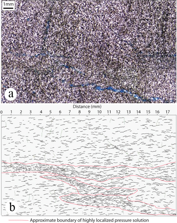
 | | Figure 2. (a) Photomicrograph showing a macroscopic PSS as seen under a petrographic miscroscope. (b) Map of micro PSS arrays within the PSS shown in (a). The thin zone (marked by red lines) is a macroscopic PSS identifiable on the hand sample and thin section slab by naked eye, which appears to be made up of a number of small PSSs localized into a tabular band-like structure. From Nenna and Aydin (2011). |
Furthermore, it is possible to identify a range of PSS zone architectures from a petrographic thin section (Figure 3) and from a cut and polished hand sample (Figure 4) both showing components of pressure solution zones, which shed light on the growth processes of pressure solution zones.
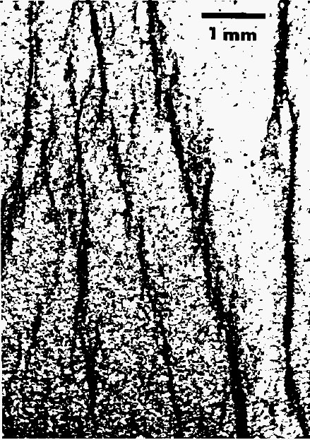
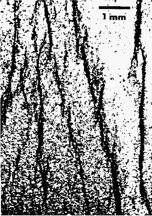 | | Figure 3. Thin section images of segmented pressure solution seams in detrital rocks of the northern Appalachian Plateau. Note that these structures were identified as 'cleavages' by the original authors. From Geiser and Engelder (1983). |
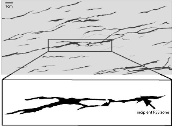
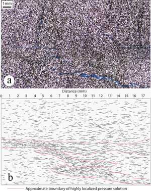
 | | Figure 4. Maps of traces of pressure solution seams on a cut and polished surface of a sample from a sandstone formation cropping out in southwest Ireland. From Nenna and Aydin (2011). Upon careful examination, it is possible to deduce that many if not only the traces are composed of laterally and transversely interconnected segments of smaller pockets of residue. This theme will be revisited in describing growth of pressure solution seams. |
Other higher order PSS zones include what is known as flaser structures (Figure 5), and what is described as lenticular zones of pressure solution seams recognized from closely spaced drill cores (Figure 6).
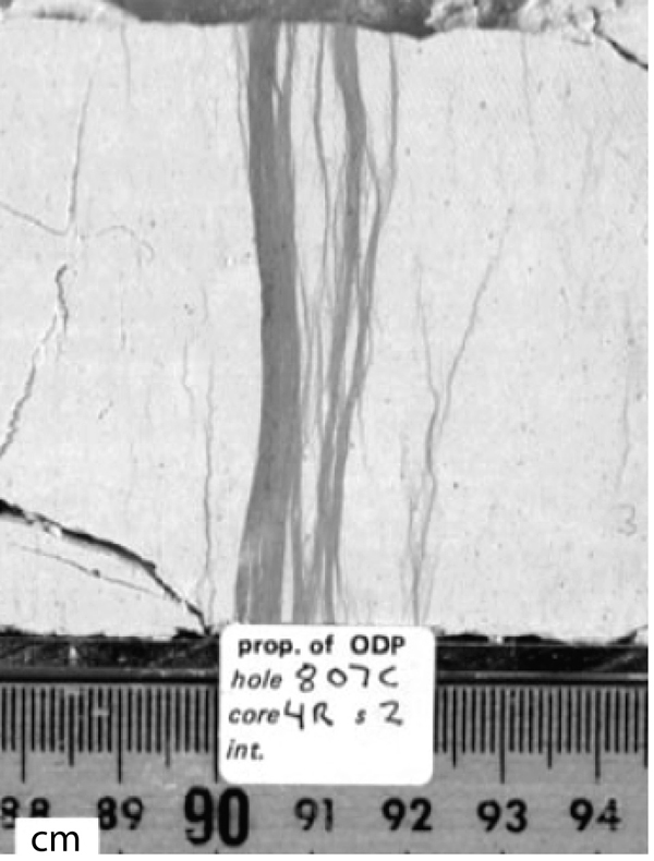
 | | Figure 5. A zone of PSS in chalk core, which is often referred to as a flaser structure. From Fabricius and Barre (2007). |
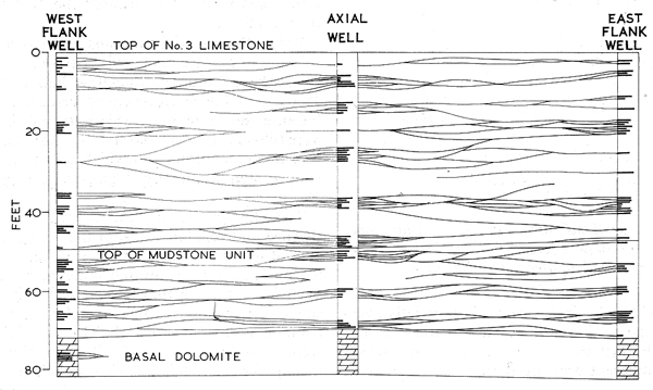
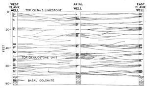 | | Figure 6. Lenticular zones of PSS as mapped from three closely spaced wells in a limestone reservoir. It was proposed that the PSS distribution was controlled by lithological variation within the unit. From Dunnington (1967). |
| |
Dunnington, H.V., 1967. Aspects of diagrnesis and shape change in stylolitic limestone reservoirs. World Petroleum Congress, 7th, Mexico, Proceedings 2: 339-352.
Fabricius, I.L., Borre, M.K., 2007. Stylolites, porosity, depositional texture, and silicates in chalk facies sediments. Ontong Java Plateau - Gorm and Tyra fields, North Sea. Sedimentology 54: 183-205, doi: 10.1111/j.1365-3091.2006.00828.x.
Geiser, P.A., Engelder, T., 1983. The distribution of layer parallel shortening fabrics in the Appalachian foreland of New York and Pennsylvania: Evidence for two non-coaxial phases of the Alleghanian orogeny. Geological Society of America, Memoir 158: 161- 175.
Nenna, F., Aydin, A., 2011. The formation and growth of pressure solution seams in clastic rocks: A field, analytical and numerical study. Journal of Structural Geology 33: 633-643, doi:10.1016/j.jsg.2011.01.014.
|